
Surgery
Feel safe and feel at ease
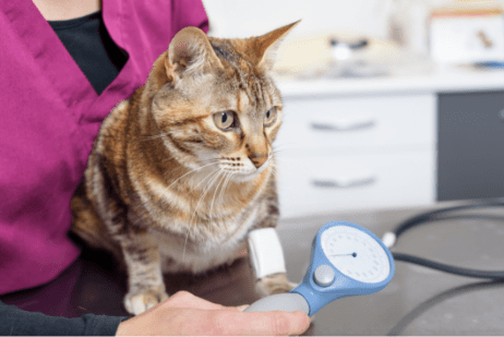
For whatever reason your pet needs surgery, this is how we want them to feel… and you too, for that matter! Surgery can be a stressful event and so we seek to minimize stress of the visit by creating a quiet environment with gentle handling, and close attention to comfort.
Before even getting to surgery our consultation gives the opportunity to discuss the diagnosis, recommended procedures, expected outcomes, and care needed. This is the time dedicated to answering your questions or addressing any concerns.
Every patient is unique.
Characteristics and health status of your pet along with factors associated with the injury or condition, may dictate different techniques for surgery. We are well equipped to deal with a wide range of these scenarios, being able to use minimally invasive techniques, fluoroscopic guidance, arthroscopy, laparoscopy or thoracoscopy.
We are a team.
Our care team is diverse, and we all share the common goal of attaining the best outcome for your pet. The network of support extends beyond the surgery department to the radiologist on site reporting on your pet’s images, veterinarians and technicians on duty observing your pet during the hospitalization period, to the cardiologist if there are any cardiac concerns. If needed, in house consults can be arranged with the other departments as well including dentistry and ophthalmology. Your pet is in good hands so you can feel safe and feel at ease.
Pulse Veterinary Specialists and Emergency
Sherwood Park 780-570-9999 *just of the Henday
Animal Surgery – Edmonton & Area
*click for directions: 450 Ordze Rd, Sherwood Park
Cruciate ligament repair & surgery for dogs & cats – Tibial Plateau Levelling Osteotomy (TPLO)
MORE INFORMATION: click here
Our Team
Originally growing up in Toronto, his love of animals and working with his hands drew him to a career in veterinary medicine. When not at the hospital Charles enjoys spending time with his family including wife Rachel and their twin daughters Charlotte and Gabrielle and their Covid puppy Jelly. An avid outdoorsman he and his family love to take advantage of the great variety of activities the area has to offer and is excited to get close(er) to the mountains!
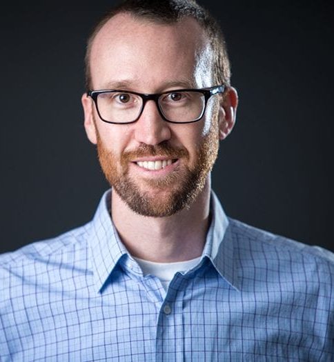
Dr. Bruce graduated from OVC in 2003 and pursued a rotating internship at the Oklahoma State University followed by a surgical internship at the Dallas Veterinary Surgical Center before returning to OVC for a small animal surgical residency. During his residency he was involved with research in spinal fractures and luxations and in the biomechanics of implant systems.
Since finishing his residency Dr. Bruce has pursued rehabilitation training through the canine rehabilitation certificate program (CCRP) through the University of Tennessee and subsequently opening and acting as director of a rehabilitation program at his previous hospital. He has trained extensively in laparoscopy, thoracoscopy and arthroscopy working as a consultant for Arthrex Vet Systems. Dr. Bruce has lectured both domestically and internationally and is looking forward to bringing his passion for veterinary medicine and his commitment to his patients to the Edmonton area.
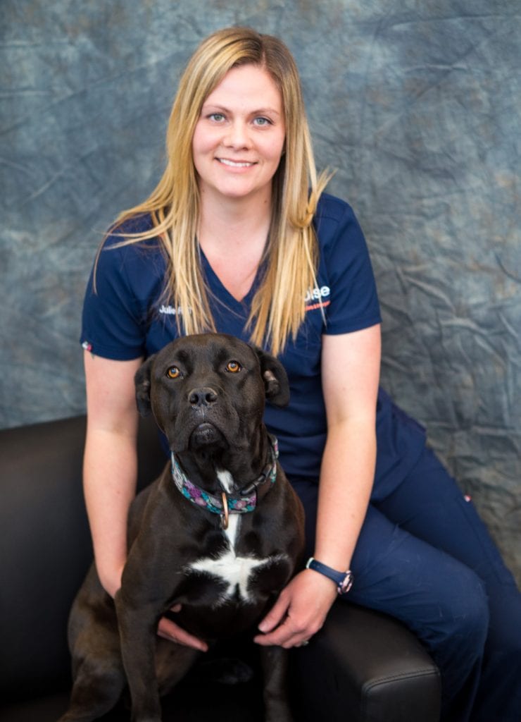
Julie graduated from the Animal Health Program at NAIT in 2006. She has been a surgery tech for over 8 years with some emergency background. Julie is hoping to complete her Veterinary Technician Specialty in anesthesia in the future. She currently resides in Red Deer with her husband, kids and two dogs, Marcus and Lyra. Julie’s interests outside of work include family time, travel, and gardening.
Bio to come

Bio to come
Bio to come
Treatments
Orthopedics includes everything from broken bones, growth deformities, congenital abnormalities, and muscular, tendon, or ligament injury.
Assessment of patients with these conditions is critical to determine what approach to take to attain the best outcomes possible. Planning may include radiographs (x-rays), and CT scans. Radiologist reporting adds to our surgical planning.
Digital images allow measurements, implant selection, and surgical planning at a very detailed level, saving time and increasing chance of success during surgery itself. During surgery all these images and surgical plans are available for viewing on our operating room viewing monitors.
Examples of orthopedic conditions that we treat along with surgical techniques:
- Cranial cruciate ligament rupture
- Tibial plateau leveling osteotomy (TPLO)
- Modified cranial closing wedge osteotomy
- Prosthetic ligament stabilization
- Fractures
- Internal plate fixation
- Interlocking nail
- External skeletal fixator
- Joint instabilities (ligament disruption e.g. Tarsal instability)
- Prosthetic ligament stabilization
- Partial or complete arthrodesis
- OCD – Osteochondritis dissecans (joint surface lesion in young dogs)
- Elbow dysplasia, joint dysplasias
- Angular limb deformities
- Patella luxation
Soft tissue surgery is a broad category of surgery for conditions such as congenital defects, respiratory abnormalities of the upper airway, thoracic (chest) conditions like lung lobe disease and vascular congenital defects, gastrointestinal conditions, urinary tract disorders, and tumors that may affect these different organ systems.
As with all surgeries, patient assessment and surgical planning is critical. CT scans are a common tool if detailed images in three dimensions are needed.
Examples of soft tissue conditions that we treat:
- Brachycephalic syndrome
- Laryngeal paralysis
- Gastric foreign bodies
- Hernias (e.g. diaphragmatic, inguinal, perineal)
- Congenital and developmental abnormalities such as liver shunts and other vascular anomalies
- Complicated mass excisions, skin flaps and grafts
- Surgical oncology involving the different organ systems
Neurosurgery in pets is most needed for intervertebral disk disease which can affect the cervical (neck) region, or along the back. We also see patients with traumatic spinal fractures and spinal area tumors.
Since neurologic impairment can take hold quickly, if there is a concern of a surgical problem, we resort to advanced imaging including CT scans. These detailed image studies are needed in almost all cases of brain or spinal disease to localize the problem and guide surgery.
Examples of neurologic conditions that we treat surgically:
- Intervertebral disc disease (IVDD) – there are some mild cases of IVDD that may not need surgery dependant on the findings of our exam.
- Vertebral fractures
- Lumbosacral disease
- Nerve root entrapment
Tools and Technology
These are invaluable to be able to accomplish what is needed during surgery. Depending on the instruments used they can decrease surgery time, improve safety, and optimize outcomes. Like all tools, they have their appropriate application.
Here is some of the equipment we have in hospital:
This is a bipolar electrosurgical device designed to deliver high current at very low voltage to tissue. It monitors tissue impedance between the jaws of the instrument and continuously adjusts the delivery of energy. The result is vessel walls that are sealed to each other. This system can be used both during open surgery and through laparoscopic portals. It is most helpful in abdominal procedures.
Scopes and cameras with 4K resolution allow visualization within the abdomen and chest through a small key-hole portal. Surgical instruments can be used under visual guidance. Vessel sealing can be used with laparoscopy and thoracoscopy.
This operating room equipment takes radiographs as still images or short video during surgery. It provides guidance for both soft tissue and orthopedic procedures. Dr. Hawkes will also use this in surgery for her cardiac procedures. Indwelling catheters, vascular accessed procedures, vascular studies, and guidance for closed (no surgical approach) orthopedic repairs are accomplished using the C-arm in conjunction with a fluoroscopy surgery table. The fluoro table is a thin carbon fibre table that doesn’t block radiographic images like conventional metallic tables.
Without going into complex biomechanics, each fixation system has its unique benefits and so it is important that different methods of fixation are available for different situations. We can perform bone plating, interlocking nail within the bone, and application of external fixators that can be used for fractures, ligament injuries, and angular limb deformities.
Like laparoscopy and thoracoscopy, 4K scopes and cameras allow visualization of more area of a joint than you can see with any given open surgical approach. With specialized arthroscopy instruments we can perform intra-articular operations such as treating a torn meniscus within the knee (stifle) or treating OCD cartilage lesions. Arthroscopy is the first step to most stifle surgeries.
Frequently Asked Questions
For most procedures there isn’t anything you need to do in advance. For all sedation and anesthesia your pet should be fasted for about 12 hours prior. Typically, this means taking away food around 10pm the night before and not feeding breakfast. Water should always be available. If your pet is on medications or any sort of home treatments, please let us know as we can give recommendations on whether they need given the morning of surgery.
This varies significantly based on the underlying reason for surgery. For elective (planned) surgeries most pets will stay for 1 night and some for 2. We will let you know our expectation for hospitalization time during our consult with you.
Pain medications are carefully selected for your pet and given before, during, and in the period after surgery. Epidurals and nerve blocks are used routinely when appropriate. Current available medications are quite good for controlling pain, but we also try to attain more comfort with enforced rest, cold compresses, and massage. In addition, we turn to short term use of mobility aids like slings or harnesses if needed. Typically, after 1 week most conditions become significantly more comfortable.
Regardless of procedure, your pet will need enforced rest for a period of time. The length of rest will be something we recommend at time of consult and discuss again when we release your pet to go home. Before your pet comes home you should find a space where your pet will be comfortable and confined. Large dog crates (this works well for cats too and will give them room for litter box, bed, and water), pens, or small rooms can work. It is important that there is not access to stairs, furniture they can jump on and off of, or doors where they could get out. They shouldn’t be following you around the house… we want them still! A leash needs to be used any time you take dogs outside for bathroom breaks…. and a leash is not a bad idea if they are at your feet near the couch. With your hand on the leash you can prevent them from running to the door and window if something suddenly caught their attention.
Providing an area to rest is about the biggest thing. Clean comfortable bedding along with supported trips outside are given as needed. Medications given on the schedule we provide to you, and monitoring of energy, appetite, and incisions are also required. Cold or warm compresses will sometimes be recommended. Pets want to lick their incisions. This can be extremely detrimental to healing and may cause infection of the underlying surgical area. E-collars or cones are used to prevent this.
If this is the case, you should call us. We are lucky the hospital is staffed with great technicians and veterinarians 24/7. They can answer your questions and give you guidance. In certain situations, they may need you to bring your pet in for a recheck. We all want to see a smooth, comfortable recovering so if there is something concerning, we would rather see them and make sure they are on track than not see them with something uncertain at home.
