
Cardiology
The importance of cardiology
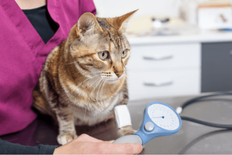
Disease of the cardiovascular system occurs in veterinary patients from young puppies to geriatric patients. It is estimated that 10 – 20% of dogs and cats have heart disease that will affect both quality and length of life. Patients with heart disease or abnormalities of the cardiovascular system benefit from early detection and diagnosis.
Our patients are important members of the family and board-certified veterinary cardiologists work with the patient, their family and their family veterinarian to help manage the patient’s heart disease and improve the patient’s quality and length of life. A veterinary cardiologist can also fix some forms of heart disease allowing a patient to live a normal life.
Our Team
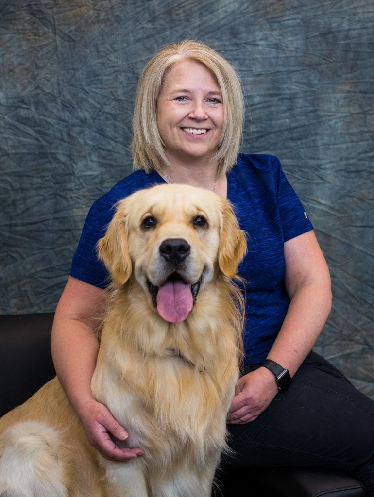
Dr. Kim Hawkes graduated from the Western College of Veterinary Medicine at the University of Saskatchewan. She completed a veterinary cardiology residency at the Ontario Veterinary College at the University of Guelph. Thereafter, she successfully obtained her board certification from the American College of Veterinary Internal Medicine—Cardiology subspecialty. Dr. Hawkes has been offering referral cardiology services for over 10 years in the Edmonton area. She is thrilled to continue offering advanced cardiology services to the Edmonton area, and continue developing relationships with the referral community.

Bio to come
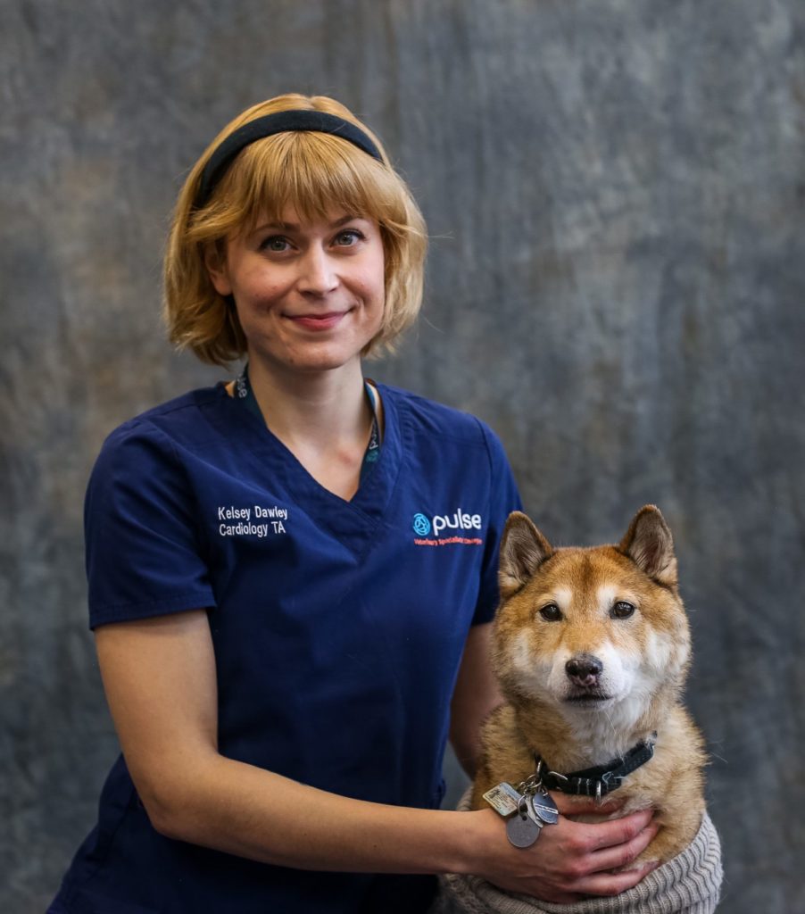

Kelsey Dawley was born and raised in Winnipeg. She attended the University of Manitoba, studying psychology (including ethology). Kelsey has been working in clinics as a veterinary assistant for over 10 years. She has a special interest in animal behaviour, and completed her Fear Free Veterinary Professional Certification. In her free time, she enjoys volunteering, hiking, camping, escape rooms, and training/adventuring with her dogs.


Bio to come
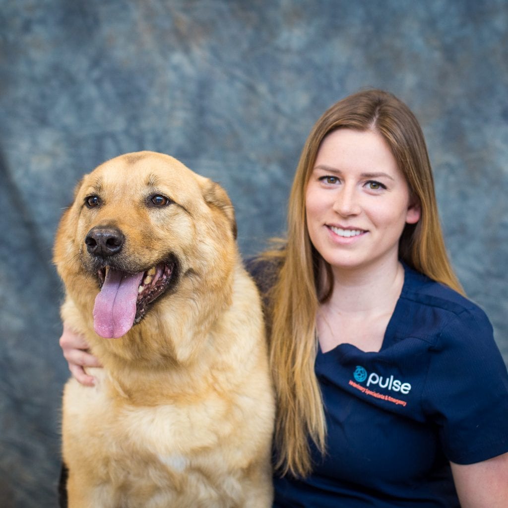

Kirsten graduated from the Animal Health Technology program at NAIT in 2018. Upon completion, she started her career in general practice and working alongside Dr. Hawkes with her mobile cardiology service. Once Pulse Vet opened in 2020, she continues to further her knowledge of cardiac diseases and management of congestive heart failure with our cardiology service. When not at work, Kirsten can be found with family and friends playing soccer, hiking, snowboarding in the winter. She also spends a significant amount of time with her very spoiled family dog, Tuke, a shepherd cross.
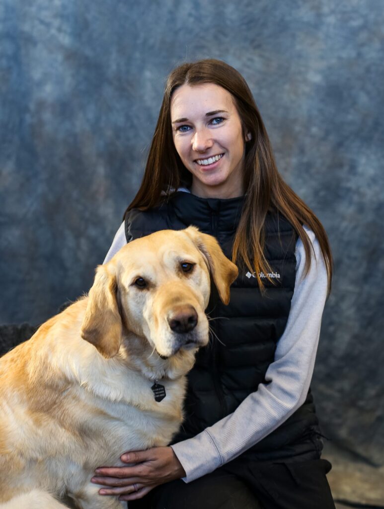

Mary graduated from the NAIT AHT program in 2021 and joined the Pulse team as a Cardiology RVT in early 2022. She finds all aspects of veterinary medicine interesting but the heart is by far her favorite! Prior to becoming an RVT, Mary obtained a Bachelor of Business Management degree from the University of Alberta and spent a few years working in accounting and human resources. Outside of work Mary keeps busy by racing motocross, going snowboarding and by playing hockey and soccer. She also loves spending time with her family and her animals at home.
Frequently Asked Questions
A veterinary cardiologist has undergone several years of advanced training and mentorship. To become a board-certified cardiologist a veterinarian must complete a minimum of a 1 year internship followed by advanced training in an approved residency training program typically of 3 – 5 years duration. After completion of their residency they attain board-certification status with successful completion of specific credentials and a long certification exam.
Board certified veterinary cardiologists focus on diagnosing and treating disease of the heart and lungs, which include:
- Congestive heart failure (CHF)
- Heart muscle disease (Dilated cardiomyopathy or hypertrophic cardiomyopathy)
- Age related changes to the valves of the heart (Degenerative mitral valve disease)
- Coughing and other breathing problems
- Congenital (present at birth) heart defects
- Cardiac arrhythmias (problems with the rate and/or rhythm of your animal’s heart)
- Diseases of the pericardium (sac surrounding the heart)
- Cardiac tumors
- High blood pressure (hypertension)
- Pulmonary hypertension (high blood pressure in the lungs)
Veterinary cardiologists will review the perform a complete assessment of the patient including a thorough physical exam, in addition to review of the patient’s history and current medications. From these initial findings, additional non-invasive tests will be discussed. Depending on the patient’s condition, diagnostic testing or treatments may include:
- Echocardiography (sonogram) – non-invasive ultrasound imaging of the heart
- Electrocardiography (ECG) – non-invasive electrical reading of the heart’s rhythm
- Blood pressure evaluation
- Holter monitor – 24 hour ambulatory ECG performed at home
- Radiography (x-rays) of the chest and lungs
- Surgical repair of congenital heart defects
- Cardiac catheterization procedures
- Balloon valvuloplasty to dilate narrowed valves
- Pacemaker implantation for animals with too slow of a heart rate
- OFA Heart Registry Certification for breeding programs
With the information gathered from the various diagnostic tests recommended your veterinary cardiologist can develop an individualized treatment plan for your companion. The veterinary cardiologist will explain their findings as well as the treatment plan with you and your family veterinarian to improve length and quality of life. Your veterinary cardiologist will also help assess anesthetic risk and help tailor anesthetic plans to increase anesthetic safety for your family companion.
Board certified veterinary cardiologists are an integral part of your animal’s health care team from the time a potential cardiac abnormality is noted. Early diagnosis and appropriate therapy of cardiac conditions helps your family companion live a longer and healthier life.
The majority of patients seen by the cardiology service are seen on an outpatient basis. The length of the appointment depends on the patient’s condition and what diagnostics are necessary. Most appointments are approximately 1 hour long. However, a patient that requires more diagnostics or a patient that is sick and having difficulty breathing may spend most of the day and night. Some patients may require hospitalization in our intensive care unit (ICU).
Echocardiograms and other cardiac diagnostics are provided on the same day as the cardiology appointments. Patients with more serious cardiac disease, or those undergoing procedures, require hospitalization in the intensive care unit where they continue to be cared for by our service.
We provide ongoing care of our patients through scheduled rechecks. Emergency services are also provided for those patients that are unstable and in need of emergency assistance and stabilization.
Undergraduate veterinary students and residents are a part of the cardiology team on a regular basis. These members of our team are involved in the appointments, diagnostic processes, and patient care under the supervision of the cardiologist. Their rotation through our service is an important part of their learning so that they can learn aspects of cardiology and prepare to help other families understand cardiovascular disease in their family companions.
MORE INFORMATION ABOUT CARDIAC DIAGNOSTICS
An echocardiogram is ultrasound imaging (sonogram) of the heart. This is a non-invasive procedure that provides real-time imaging of the heart to evaluate heart size and function. The complete exam typically involves 2D and Doppler imaging. The 2D imaging allow us to evaluate structure and function (contraction and relaxation) of the heart in real-time. Colour Doppler allows us to follow the flow of blood through the heart, identifying any leaky valves or turbulent blood flow. Spectral Doppler allows the the measurement of speed of blood flow through the heart and this can allow us to then estimate cardiac pressures. Additional advances in the field of echocardiology include 4D echo as well as tissue Doppler imaging and speckle tracking. These advances allow board-certified veterinary cardiologists to assess heart structure and function in even more detail. The cardiology service at Pulse Veterinary Specialists & Emergency uses the newest and most advanced echocardiography unit the Vivid E95 in hospital and the Vivid iq for portable cardiac ultrasound.
An electrocardiogram (ECG) is used to assess a patient’s heart rhythm. If an abnormal heart rhythm (arrhythmia or dysrhythmia) is noted from your veterinarian or during our exam we will recommend further assessment with an ECG. This allows us to assess the electrical activity of the heart. Animals with heart disease can commonly have secondary arrhythmias such as Atrial Fibrillation or ventricular ectopy. The ECG can also help us assess response to therapy.
We use a Philips TC70 ECG which allows us to assess multiple simultaneous leads with a real time ECG displayed on a large screen.
A Holter monitor is a small, lightweight ambulatory electrocardiogram (ECG) that records 24 to 48 hours of heart rate and rhythm data. This allows us to obtain more complete disclosure on the patients rhythm and assess for intermittent disturbances of the cardiac rhythm. Ambulatory ECG (AECG) is recommended in patients with dysrrhythmias and also in patients that have a history of syncope (fainting). The Holter is also used to assess response to anti-arrhythmic therapy for a variety of arrhythmias.
Some Holters can stay on for a longer period of time (days) – these are best for intermittent episodes that may not occur daily. This is called an event monitor and requires the pet owner to trigger the device following an episode.
Telemetry is used for some in hospital patients to allow us to continually monitor heart rhythm and other patient parameters while in hospital.
Free fluid can accumulate in the chest and abdominal cavities of some patients with heart disease. Fluid in the chest (pleural effusion) and abdomen (ascites) can be the result of heart failure and can cause difficulty breathting and discomfort. Removal of the fluid is called centesis (thoracocentesis and abdominocentesis), and this will improve clinical signs and comfort in patients with CHF. Medical therapy will then try to slow down or delay the recurrence of the fluid accumulation. In some animals a cardiac tumour can bleed and cause a build-up of fluid around the heart called pericardial effusion. The pericardial effusion can result in a constraining effect on the heart and compromise of the circulatory system. Therefore removal of the fluid from the pericardial sac (pericardiocentesis) is required in those patients that are experiencing this degree of compromise (cardaic tamponade). We routinely perform any required centesis procedure at Pulse Veterinary Specialists & Emergency. In some cases a sample of the fluid will be submitted to the veterinary lab for further assessment.
Thoracic radiographs are x-rays of the chest. We routinely take 3 different views of the chest to assess for build up of fluid in the lung tissue (pulmonary edema). When the heart (the pump) fails due to some conditions (eg. myxomatous mitral valve degeneration) there is a build up of volume and pressure in the capillary bed of the lungs resulting in the leakage of fluid into the lung tissue. This accumulation of fluid in the lung tissue results in exercise intolerance, difficulty breathing and coughing. Thoracic radiographs allow us to assess the presence and severity of pulmonary edema. They also allow us to assess for pleural effusion, respiratory disease such as chronic bronchitis or neoplasia (cancer).
Measuring blood pressure is an important part of a cardiac examination in any animal – that’s why we include it with all of our cardiac evaluations! Systemic hypertension is common in animals with many affected animals showing no signs of disease.
Hypertension can have a negative impact on the heart function in animals with heart disease. Therefore addressing hypertension is an important part of a treatment plan in any animal with heart disease.
Some of the medications prescribed to a patient with heart disease can lower the blood pressure – therefore, trending blood pressure becomes an essential part of monitoring therapy in these animals.
Many veterinarians use Doppler blood pressure to measure the pressure indirectly. A small ultrasonic crystal is used to hear the arterial pulse while a small cuff is inflated and deflated to allow measurement of the systolic blood pressure. Oscillometric blood pressure monitors are also used. In animals, the systolic blood pressure is very similar to that in a human.

