Diagnostic Imaging
What is Diagnostic Imaging?
Our patients can’t tell us where it hurts. This is why the Diagnostic Imaging department at Pulse Veterinary Specialists and Emergency offers the most state-of-the-art imaging equipment to help ‘look inside’ your pet, aiding in diagnosis of illness. Diagnostic Imaging is a subspecialty of veterinary medicine that focuses on using advanced imaging technologies, such as x-rays, ultrasound, CT, and MRI, for the diagnosis and management of complex cases.
A specialist in diagnostic imaging is called a “radiologist”, a call-back to times when the only type of imaging available to veterinary patients was x-rays or radiographs. Thankfully, many advances in imaging from the human medical field have made their way to veterinary medicine, including ultrasound, CT, and MRI. Pulse Veterinary Specialists and Emergency is excited to make these imaging modalities available to the pets of Alberta’s capital region! Click on a modality below to learn more about how it can help with diagnosis in your pet.
Our board-certified veterinary radiologist, Dr. Lukas Kawalilak, is an expert in all areas of diagnostic imaging and works closely with Pulse clinicians and your primary care veterinarian in developing an imaging plan that is best for each individual patient’s health needs. Dr. Kawalilak’s specialized training and years of experience allows him to see abnormalities and clarify findings from diagnostic imaging procedures. This is key to providing the best possible care for your pet.
Our Team

Dr. Lukas Kawalilak graduated from the Western College of Veterinary Medicine in 2013. He completed a veterinary diagnostic imaging residency in 2018 that included training at both the Michigan State University and Colorado State University. He successfully completed board certification in Veterinary Radiology in 2018. He is looking forward to working with the referral community as the first veterinary radiologist in Northern Alberta.
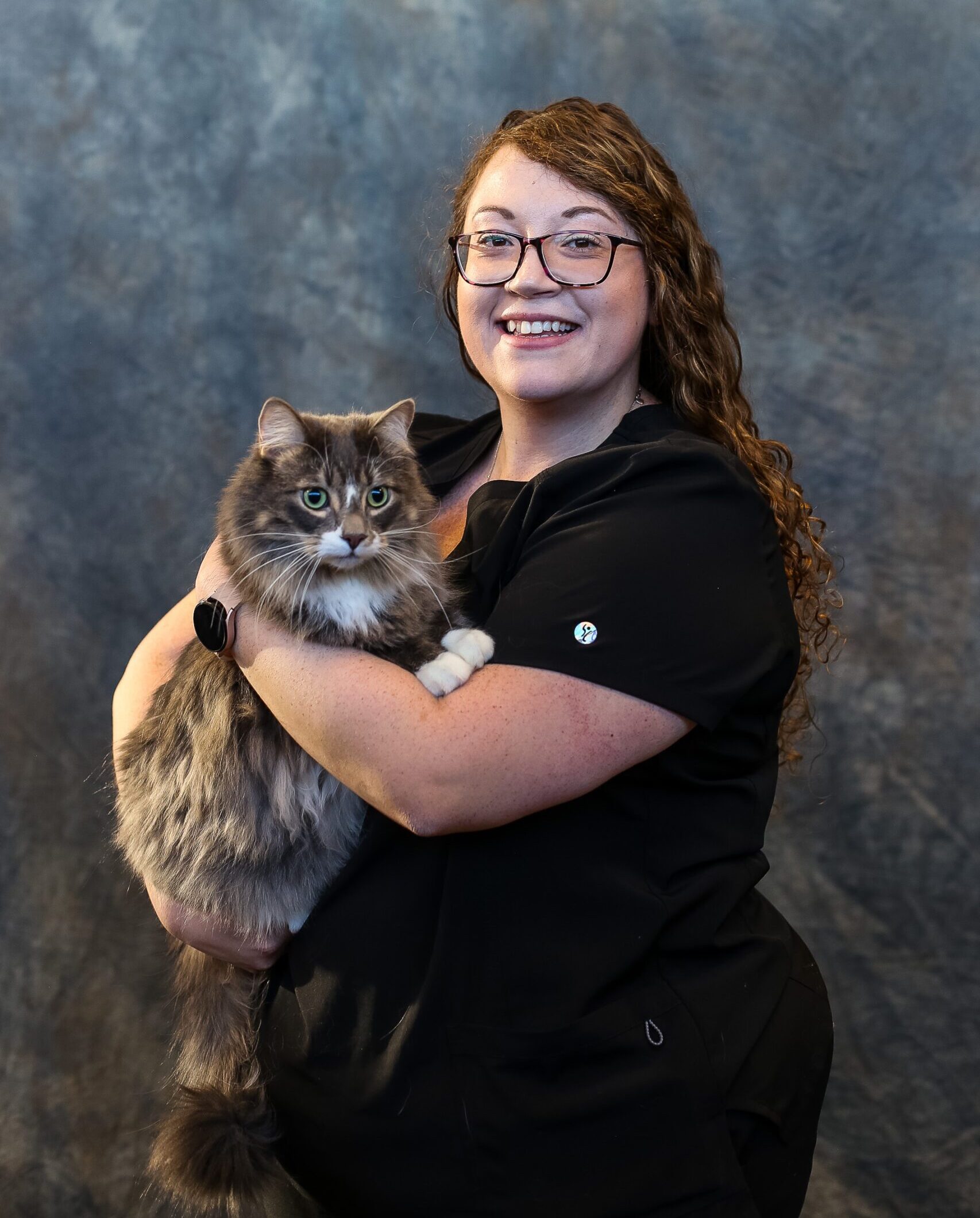
Born and raised in Prince Edward Island. Leah moved to Edmonton, Alberta in 2013 to pursue her love for animals. Leah graduated from NAIT Animal Health Technology program in 2019. She spent the last couple years in general practice with experience in small animal and exotics before joining the Pulse team. In her free time, she likes to play with her two cats (Moose and Chloe), reading, and spending time outside with family and friends.
Imaging Modalities Available at Pulse Veterinary Specialists and Emergency
The Diagnostic Imaging department at Pulse Veterinary Specialists and Emergency offers the most state-of-the-art imaging equipment to help in diagnosing your pets illness.
What we offer:
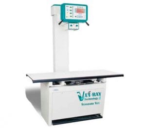
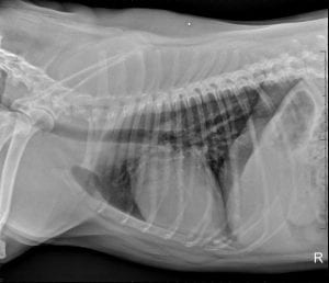
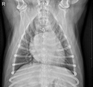
Radiography is the oldest and most common diagnostic imaging tool and is the first step in obtaining a diagnosis for pets. We offer state-of-the-art digital radiography suites, which are able to provide a rapid acquisition of digital images that are clearer than conventional film radiographs. These images can also be manipulated on a computer, allowing our doctors to view them in ways not possible with films. All digital images are stored electronically, allowing for easy retrieval of patient images on computers throughout the hospital, as well as easily transferred to you or your primary care veterinarians for review
Clinical conditions and procedures where referral to Pulse for x-ray imaging may be considered (but not limited to):
- pneumonia and other lung problems
- heart failure and other heart problems
- cancer
- intestinal obstruction
- causes of pain and limping
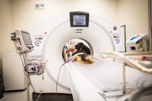
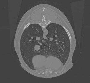
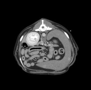
Computed Tomography (CT) is a non-invasive diagnostic imaging technique in which X-rays are taken 360° around the patient to obtain cross-sectional images or ‘slices’ of the body. This makes a CT scan one of the best tools for examining the chest or abdomen of a pet. A CT machine utilizes a dedicated high-speed computer to produce images and allows the images to be manipulated to specifically enhance certain types of tissues, according to what area of the body is being examined. The computer also allows three-dimensional reconstruction of images that can help our clinicians localize disease and accurately plan procedures such as surgery. Our high-speed multidetector CT machines allow detailed exams to be performed in a very short period of time, minimizing the amount of time a patient needs to be sedated or anesthetized.
Clinical conditions and procedures where referral to Pulse for CT imaging may be considered (but not limited to):
- cancer or other diseases of the nasal cavity and lungs
- abnormal bloods vessels in the liver
- masses arising from internal organs such as the spleen
- disorders of the abdomen such as cancer or kidney stones
- disorders of bones and joints such as elbow dysplasia
- guide biopsies and other minimally invasive procedures
- plan surgery
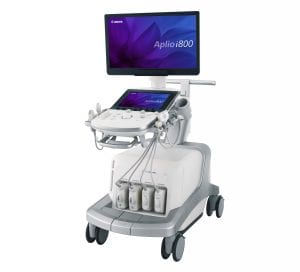
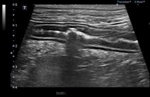
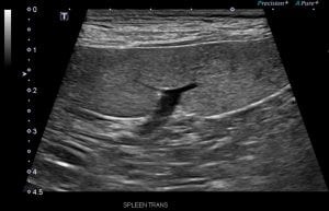
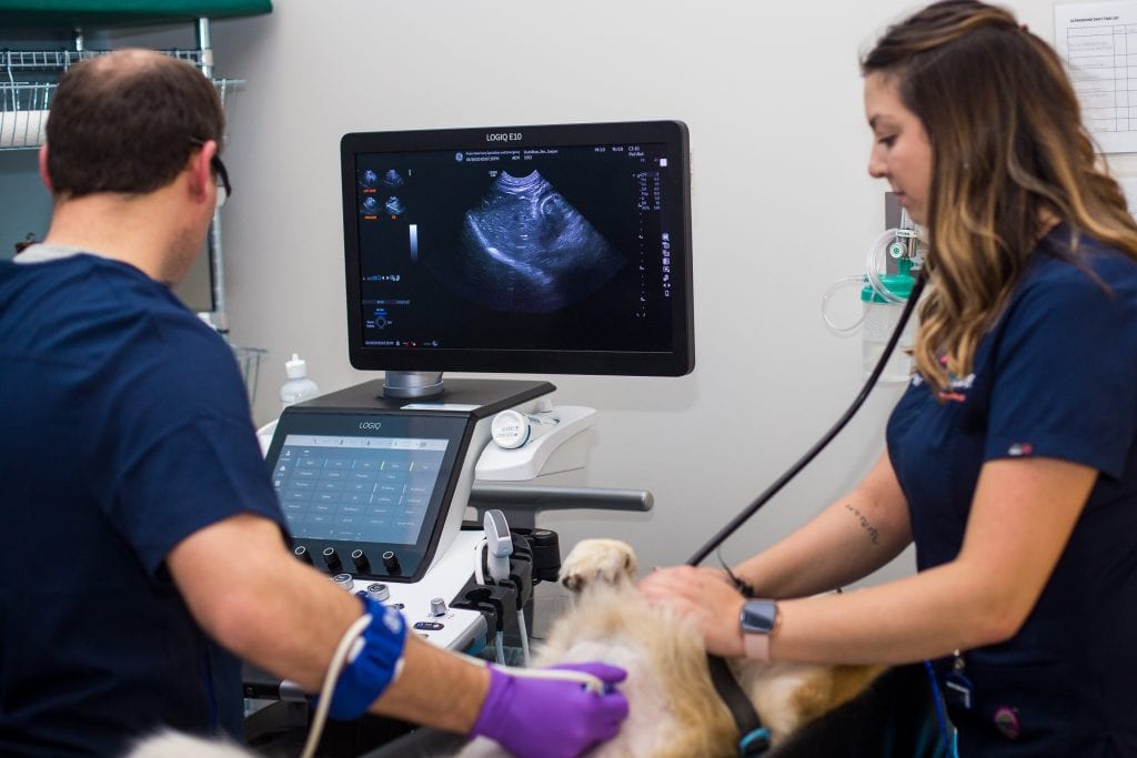
Utrasonography provides a painless and non-invasive method of evaluating the internal structures of the body and providing a view of abnormalities that cannot otherwise be seen with other diagnostic methods. Ultrasound allows for evaluation of internal organs, musculoskeletal injuries, abnormalities within the gastrointestinal system, as well as fluid or masses within the body. Together, with patient medical history and a thorough physical exam, an ultrasound exam can often diagnose the source of pain or illness. Ultrasound is also an excellent tool to obtain tissue samples without putting your pet through an invasive surgery. These procedures known as a ‘fine needle aspirate’ or ‘core biopsy’ take a sample of the tissue in the area that appear abnormal and can then be examined by a veterinarian or sent to the lab for analysis.
Clinical conditions and procedures where referral to Pulse for ultrasound imaging may be considered (but not limited to):
- abdominal pain
- pancreatitis
- enlarged abdominal organ
- stones in the kidney or bladder
- portosystemic shunts
- needle biopsies in which needles are used to extract a sample of cells from organs for laboratory testing
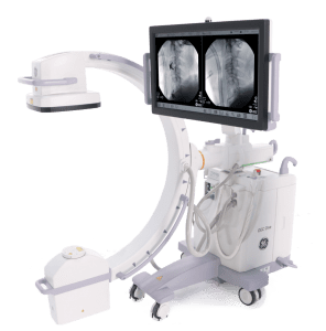
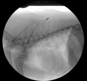
Fluoroscopy is a continuous series of very low dose x-ray images that let veterinarians see live images of the inside of the body in motion. This dynamic evaluation of a patient, allows us to image a region of interest continuously, while simultaneously watching on the screen. The machine that generates these images is called a C-arm and can be rotated around the patient, allowing images to be obtained from any angle without moving the patient. For example, fluoroscopy of the chest allows us to watch the heart beat and the lungs expand.
Clinical conditions and procedures where referral to Pulse for fluoroscopic imaging may be considered (but not limited to):
- swallowing disorders such as megaesophagus or esophageal stricture (narrowing)
- airway diseases such as collapsing trachea
- motility problems of the stomach and intestines
- urinary diseases such as obstructions including stone or strictures
- flow of blood through abnormal vessels
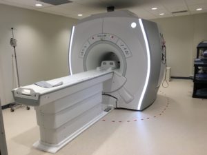
MRI is a powerful imaging technique that utilizes a super-conducting magnet inside a doughnut-shaped scanner that looks similar to a CT scanner. MRI and CT are very different though! When the patient is placed inside the MRI scanner, their protons align to the magnetic field. Throughout the scan, radiofrequency pulse sequences affect the magnetic field and the protons realign; these changes are detected using coils wrapped around the area being imaged. This means no radiation is used when a patient is imaged using MRI.
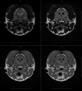
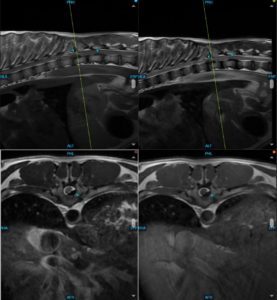
MRI is the imaging tool most often recommended by veterinary neurologists and radiologists to evaluate the nervous system. In contrast to CT, which is better for bone evaluation, MRI is significantly better at imaging soft tissues, such as the brain, spinal cord, and intervertebral discs. As any movement blurs the images dogs and cats must be placed under general anesthesia for the MRI scan. Once safely under anesthesia, the procedure usually takes between 45 minutes and 2 hours to complete depending on the region of the body being scanned. During this time, your pet is closely monitored by a veterinary technician. Since your pet is under anesthesia for the procedure, we have to focus our scan on the region of interest for patient safety, which is why “whole body” MRI is not performed at Pulse.
Examples of diseases affecting the brain and spinal cord diagnosed using MRI include:
- Intervertebral disc disease (herniation and protrusion)
- Fibrocartilagenous embolism
- Inflammatory – immune mediated or infectious meningoencephalitis
- Brain tumors
- Stroke
- Peripheral nerve sheath tumor
Frequently Asked Questions
Because of radiation safety regulations, owners cannot be present in protected areas during the study. Rest assured, your pet will be well cared for by our registered animal health technologists and animal care assistants
Many x-ray and ultrasound examinations can be done with gentle manual restraint.
- However, sedation or anesthesia is used for pets who are excitable, when samples of organ are taken (fine needles aspirates or biopsies), or for imaging studies that have to be very carefully positioned for surgical planning.
- A short-acting anesthetic or sedative may be needed to help relax your pet during the procedure and prevent potential complications.
CT and MRI examinations must be performed under general anesthesia to get high quality images for interpretation.
- However, the duration of anesthesia is generally short, and the patient is monitored using sophisticated equipment including electrocardiogram, blood pressure and respiratory monitoring devices.
- Pulse uses modern anesthesia drugs that allow for quick onset of and recovery from anesthesia. Your pet may still be a little drowsy the evening following its imaging procedure.
The dose of radiation used to take x-rays or CT images are very low and will not cause harm to your pet.
Pets having an ultrasound or CT exam should ideally not eat for twelve hours prior to the procedure. Please continue to provide free access to fresh water.
- The presence of food in the stomach makes it nearly impossible for the ultrasound beam to penetrate to the organs to be studied. Even if the animal has only a small meal or a cookie, he or she may swallow gas with it, which will block the ultrasound beam.
- For CT exams, having food in the stomach can increase the risk of vomiting following sedation or anesthesia; an empty stomach can help prevent this complication
Pets having x-rays performed usually do not need to be fasted prior to their imaging, but check with your Pulse veterinarian to confirm
Ask your veterinarian for instructions if your pet is on any medications prior to fasting.
Since ultrasound waves are not transmitted through air, it is imperative that the hand-held probe makes complete contact with the skin. This means that the patient’s fur must be shaved to perform an ultrasound examination.
Without shaving, the imaging obtained by the ultrasound machine are not useful in diagnosing disease
Our dedicated diagnostic imaging staff will do their best prepare your animals skin with utmost care. There may be some mild redness and itching after the ultrasound exam from the gel and alcohol used to increased probe contact with the skin.
Fine needle aspirates (FNA) and core needle biopsies are methods of collecting cells or tissue from a specific site such as an organ (kidney or liver), an undetermined mass, or area to look for signs of infection, cancer, or other conditions.
- An FNA uses a smaller needle and is less painful, but acquires a smaller amount of tissue for analysis
- A biopsy uses a larger needle and obtains a larger sample, but requires the animal to be heavily sedated
The radiologist inserts a needle or biopsy instrument into the area and withdraws a sample of tissue. The material is then examined under a microscope or sent to the lab for further analysis.
An FNA or biopsy are often needed to determine if tissue that appears abnormal on imaging is diseased or normal
FNAs and biopsies can be guided using ultrasound or CT
It is best if your pet arrives to their appointment with a full bladder so that we have the best opportunity to perform a thorough ultrasound.
If this is not possible your pet may have to board for a few hours to give time for the bladder to fill back up.
Contrast containing iodine is given intravenously during most CT and some fluoroscopy exams. It is given to patients to help determine the underlying cause for the patient’s illness. Contrast is critical to distinguishing normal from abnormal structures in parts of the body such as the lungs, liver, spleen, and bones.
Contrast in veterinary patients is very safe. Reactions to contrast agents used in CT are rare in veterinary medicine. Each patient is closely monitored for any potential reactions to the injection. Veterinary patients undergoing imaging that receive contrast during the procedure do so under the guidance of trained animal health technologists and doctors.

