

Neurology
What is Neurology?
Neurologists manage diseases that affect the brain, spinal cord, or peripheral nerves and muscles. The nervous system is involved in generating movements of the body, vision, balance, hearing, and touch/sensation. It also helps control urination and defecation, and plays a role in regulation of intestinal motility and vital parameters. With so many normal daily functions governed by the neurologic system it is no wonder that loss of these functions can be serious and can cause a wide variety of signs. Some conditions can come on very suddenly whereas others have a more gradual progression. Often times, even conditions we expect to have a gradual progression, can present acutely as the nervous system is very good at compensating until it reaches a threshold. Neurologic disease can be related to immune mediated disease, infection, congenital defects, tumors/cancer, trauma, degenerative disease, or secondary to some metabolic conditions or toxins.
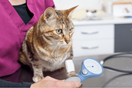
The first step in diagnosing a neurologic condition is to localize where the problem is coming from. One of the highly tuned skills of a veterinary neurologist is determining which portions or tracts of the nervous system are affected, whether it is diffuse or from a specific location. This is done by a thorough neurologic examination that assesses mentation, cranial nerves, gait analysis, paw placement proprioception testing, spinal reflexes, and assessing for pain. Based on the patients history, presenting symptoms, and neurolocalization, a list of possible diseases is determined. Depending on where the problem is localized to, along with the presenting signs, further tests can be performed to reach a diagnosis. Because many diseases can affect an area of the nervous system, and symptoms are a reflection of the area affected, further testing is needed to obtain a definitive diagnosis. Often, this involves advanced imaging (MRI, CT, or sometimes a combination), spinal tap, or electrodiagnostics (electromyography, nerve conduction) and biopsies. Depending on the diagnosis, neurologic diseases can be treated with medication, surgery, rest, and rehabilitation therapy or a combination of these treatments.
Pulse Veterinary Specialists and Emergency
Sherwood Park 780-570-9999 *just of the Henday
*click for directions: 450 Ordze Rd, Sherwood Park
Our Team
Dr. Casey Smith graduated from the University of Glasgow School of Veterinary Medicine. After graduation, she spent a year at a rotating internship in Pittsburgh followed by a surgery/neurology internship in San Antonio. She then returned to Canada to pursue a neurology/neurosurgery internship in Vancouver. She completed a 3-year residency in neurology/neurosurgery at the University of Wisconsin-Madison and became board certified in Neurology with the ACVIM in 2021.
Her professional interests include brain and spinal surgery, breed related neurologic disease, movement disorders, EEG, and MRI.
Dr. Smith is a French Bulldog fanatic with a trio of the mischief makers also “employed” in the Neurology department – Ruby (the Queen B), Arthur (just here for the vibes and snacks), and Midge (chaotic wild child). She is a Harry Styles enthusiast, and a collector of far too many houseplants, more books than she can keep up with, and vintage estate sale finds.

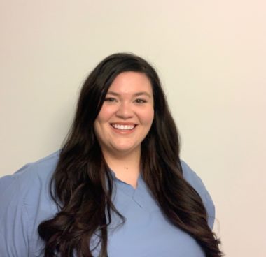
Bio to come.

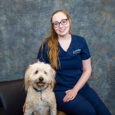
Bio to come
Bio to come
Bio to come
Common conditions
Diseases affecting the brain can have a variety of signs depending on which area(s) of the brain are involved as each main area has a different function. Some conditions only involve a focal area whereas other diseases (meningoencephalitis, for example) can affect multiple parts of the brain.
Common signs of intracranial disease can include:
- Seizures
- Behaviour change or loss of normal tricks/commands
- Altered mentation/level of alertness
- Circling
- Weakness
- Incoordination
- Balance issues
- Vision changes
- Pacing
- Cranial nerve deficits (changes in the sensory or motor
function of nerves going to the head/face) - Head tilt
- Abnormal eye movements (nystagmus)
- Tremors
Examples of intracranial diseases diagnosed and treated by neurologists:
- Idiopathic epilepsy
- Inflammatory – immune mediated or infectious meningoencephalitis
- Vestibular disease
- Hydrocephalus
- Caudal occipital malformation syndrome (COMS)
- Brain tumors
- Stroke
- Head trauma
- Cognitive dysfunction
- Tremors
Diseases affecting the spinal cord can affect multiple areas or, more commonly, one specific region. This can be anywhere from the neck to the tail and can have a variety of underlying causes.
Common signs of spinal cord disease can include:
- Pain
- Abnormal posture
- Weakness or paralysis in one or multiple limbs
- Incoordination
- Scuffing or dragging paws
- Changes to urinary or fecal continence
- Changes to reflexes and muscle tone in the limbs or tail
Examples of diseases affecting the spinal cord diagnosed and treated by neurologists:
- Intervertebral disc disease (herniation and protrusion)
- Fibrocartilagenous embolism
- Acute non-compressive nucleus pulposus extrusion (ANNPE)
- Discospondylitis
- Steroid responsive meningitis arteritis (SRMA)
- Other immune mediated or infectious meningomyelitis
- Spinal tumors
- Atlanto-axial instability
- Spinal subarachnoid diverticula
- Vertebral malformations
- “Wobbler’s syndrome” (cervical spondylomyelopathy)
- Degenerative myelopathy
- Lumbosacral stenosis
- Syringomyelia
Common signs of neuromuscular disease can include:
- Generalized weakness
- Exercise intolerance and collapse
- Changes to reflexes and muscle tone
- Muscle atrophy
- Abnormal sensation
- Painful muscles
- Reduced range of motion of limbs
Examples of peripheral/neuromuscular diseases diagnosed and treated by neurologists:
- Myasthenia gravis
- Masticatory muscle myositis
- Botulism
- Tick paralysis
- Acute polyradiculoneuritis (“Coohhound paralysis”)
- Facial paralysis
- Trigeminal neuropathy (idiopathic, neuritis, tumor)
- Degenerative polyneuropathy
- Brachial plexus injury
- Peripheral nerve sheath tumor
Frequently Asked Questions
No. The neurologic examination itself is not painful. Some dogs/cats don’t like the part of the examination where their paws need to be touched but this is usually short lived and generally doesn’t cause a lot of stress. Imaging tests like MRI and CT scan are not invasive and are performed under general anesthesia. Spinal tap is also performed under anesthesia and electrodiagnostics are performed under heavy sedation or anesthesia and so there is no discomfort involved in these procedures.
This depends on the region of the nervous system that is affected. In general, neuromuscular diseases don’t require an MRI for diagnosis however for brain or spinal cord diseases it is the best way to evaluate all of the structures involved. It provides detail of soft tissue structures that CT or radiographs can’t.
A spinal tap is a procedure that collects the fluid that covers the brain and spinal cord. In dogs and cats we can collect from a spot behind their skull (cerebellomedullary cistern) or the lower lumbar spine, sometimes both. Because this fluid bathes the central nervous system it can give more information at a cellular level of what is going on especially regarding inflammation. It also helps complement findings on MRI and can
help us obtain a definitive diagnosis. In general, spinal tap is performed after advanced imaging to ensure that the spinal tap is necessary (as not all conditions require this for diagnosis). In particular with brain diseases, the MRI helps us ensure that spinal tap is safe to perform.
For people it is easy to say “you need to stay still for this MRI/CT”. For dogs and cats, this isn’t so easy. Advanced imaging takes longer to obtain the images than it does for an x-ray, for example. To obtain all of the scans needed, and have an exact comparison between different types of sequences used, our patients need to stay perfectly still and in the same position for the duration of the MRI/CT. Therefore, full general anesthesia is needed for MRI.